Images for “Vampirism”:

Image used from: https://www.livescience.com/40843-real-dracula-vlad-the-impaler.html
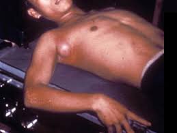
Image used from: here

Image used from: here

Image used from: here

Image used from: here. Side note, this is a different image from the one referenced in the episode. I showed Mia a cartoon image of spinal tuberculosis in the episode.

Image used from: https://www.livescience.com/40843-real-dracula-vlad-the-impaler.html

Image used from: here. This is a gram stain of Y. pestis.

Image used from: here

Image used from: here. This is an acid fast stain of M. tuberculosis.

Image used from: https://www.howrahtb.com/indoor.html. Side note, the image on the right is her on the day of her discharge from the hospital. She did recover.
Images for Human Blood:

Image used from: Humours


Image used from: https://courses.lumenlearning.com/suny-ap2/chapter/erythrocytes/

Image used from: https://www.ahajournals.org/doi/full/10.1161/CIRCRESAHA.119.314977

Image used from: Blood

Image used from: https://courses.lumenlearning.com/suny-ap2/chapter/erythrocytes/

Image used from: https://www.youtube.com/watch?v=7aCeaI9nPsg

Your Homework: used from here
Images for Electricity Part 2:

Image used from: https://www.livescience.com/53509-faradays-law-induction.html

Image used from: https://www.livescience.com/53509-faradays-law-induction.html

Image used from: https://tjregister.wordpress.com/2017/03/14/lenzs-law-activity/

Image used from: https://en.wikipedia.org/wiki/Homopolar_generator

Image used from: https://brainly.com/question/9213822


Image used from: https://www.youtube.com/watch?v=Ylgb8FFMgd4

Image used from: here


Images for Electricity Part 1:


Image used from: https://www.slideserve.com/eliot/chapter-10
This image doesn’t really have anything to do with with the episode, but Mia wanted you the people to see it.

Image used from: https://circuitideas311.weebly.com/lesson-one/chapter-one

Images for Nuclear Fission

Image used from:https://www.atomicheritage.org/profile/lise-meitner
Physicist Lise Meitner

Image used from: https://www.centrusenergy.com/learn-more/uranium-enrichment/gaseous-diffusion/
Gaseous diffusion


Image used from: https://kindgardensupply.com/products/ai-5-liter-glass-separatory-funnel-kit-with-all-ptfe-valves
Separatory funnel in action

Image used from:https://www.livescience.com/37206-atom-definition.html

Image used from: https://www.osha.gov/SLTC/radiationionizing/background.html
Radiation particles and penetration levels.

Image used from: http://www.hk-phy.org/energy/power/nuclear_phy04_e.html
Fission chain reaction

Image used from: https://www.dummies.com/education/science/chemistry/the-basics-of-nuclear-fission/
Plutonium 239 from Uranium 238 reaction

Image used from: https://www.atomicheritage.org/history/science-behind-atom-bomb

Image used from: https://science.howstuffworks.com/nuclear-reactor.htm
Images for Innate Immunity Part 4

Image used from: The Immune System 4th Edition by Peter Parham. Image depicts the pentameric C-reactive protein.

Image used from: The Immune System 4th Edition by Peter Parham. Image depicts mannose binding lectin complex.

Image used from: The Immune System 4th Edition by Peter Parham.

Image used from: The Immune System 4th Edition by Peter Parham.

Image used from: The Immune System 4th Edition by Peter Parham. Image depicts C1 component from classical complement pathway.

Image used from: The Immune System 4th Edition by Peter Parham.

Image used from: The Immune System 4th Edition by Peter Parham.

Image used from: The Immune System 4th Edition by Peter Parham.

Image used from: The Immune System 4th Edition by Peter Parham.

Image used from: The Immune System 4th Edition by Peter Parham.
Images for Innate Immunity Part 3

Image used from: The Immune System 4th Edition by Peter Parham.

Image used from: https://www.frontiersin.org/articles/10.3389/fphar.2020.00554/full

Image used from: The Immune System 4th Edition by Peter Parham.

Image used from: The Immune System 4th Edition by Peter Parham.

Image used from: The Immune System 4th Edition by Peter Parham.

Image used from: The Immune System 4th Edition by Peter Parham.

Images for Innate Immunity Part 2

Image used from: The Immune System 4th Edition by Peter Parham

Image used from: The Immune System 4th Edition by Peter Parham

Image used from: The Immune System 4th Edition by Peter Parham

Image used from: The Immune System 4th Edition by Peter Parham

Image used from: The Immune System 4th Edition by Peter Parham

Image used from: https://iai.asm.org/content/87/10/e00127-19

Image used from: The Immune System 4th Edition by Peter Parham

Image used from: The Immune System 4th Edition by Peter Parham
Images for Innate Immunity Part 1

Image from: The Immune System 4th Edition by Peter Parham.

Image from: The Immune System 4th Edition by Peter Parham.

Image from: The Immune System 4th Edition by Peter Parham.

Image from: https://www.mun.ca/biology/scarr/Gr10-32.html

Image from: The Immune System 4th Edition by Peter Parham.

Image from: The Immune System 4th Edition by Peter Parham.

Image from: The Immune System 4th Edition by Peter Parham.
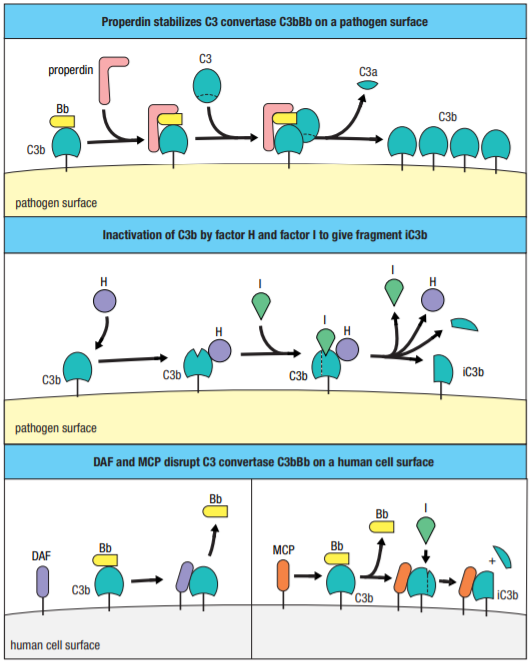
Image from: The Immune System 4th Edition by Peter Parham.

Image from: The Immune System 4th Edition by Peter Parham.

Image from: The Immune System 4th Edition by Peter Parham.

Image from: The Immune System 4th Edition by Peter Parham.
Image for Drug Development Part 2

Image used from: https://abouquetfrommendel.wordpress.com/2012/02/21/bitter-but-beneficial-salicylic-acid-and-willow-bark/
This image depicts the chemical structures for salicin (the active ingredient in willow bark), salicylic acid (a refined version of salicin), and acetylsalicylic acid, which is modern day aspirin.
Images for COVID-19 Part 2 of 2

Image used from: https://www.livescience.com/covid-19-double-lung-transplant.html
CT scan of COVID-19 patient’s lungs with extensive tissue damage (ground glass opacity).

Image used from: here
This image depicts the three main steps of PCR, and how the probe is annealed to the genetic sequence.

Image used from: https://newsroom.uw.edu/news/covid-19-coronavirus-spike-holds-infectivity-details
This is a three-dimensional depiction of a SARS-CoV-2 spike glycoprotein.

Image used from: https://www.livescience.com/covid-19-double-lung-transplant.html
Lung from COVID-19 patient who received double lung transplant.

Image used from: https://www.asbmb.org/asbmb-today/science/031720/what-makes-remdesivir-a-promising-antiviral
On the left is the chemical structure for remdesivir, the RNA adenosine analog being used as an antiviral. On the right is the chemical structure for an actual RNA adenosine nucleotide.
Images for Ep. 1: Covid-19 Part 1

Image used from https://www.sciencedirect.com/science/article/pii/S2090123220300540#b0265
Crude cartoon of SARS-CoV-2 virion. The genome and structural proteins are all represented in the image.

Image used from https://www.sciencedirect.com/science/article/pii/S2090123220300540#b0265
This image depicts the single strand of genomic RNA for both SARS-CoV and SARS-CoV-2. The genome has been broken up and labeled to show what sequences code for what proteins.

Image used from https://commons.wikimedia.org/wiki/File:0302_Phospholipid_Bilayer.jpg
This image depicts a phospholipid bilayer. This is what the membranes of our cells are made from.

Image used from https://www.sciencedirect.com/science/article/pii/S2090123220300540#b0265
This image depicts a simplified, but comprehensive replication cycle for SARS-CoV-2.

Image used from https://www.sciencedirect.com/science/article/pii/S2090123220300540#b0265
This image shows how the different genera of coronaviruses use different hosts. Notice, beta-coronaviruses are capable of causing disease in humans. MERS, SARS, and SARS-Cov-2 are all beta coronaviruses.

Image used from http://www.healthandfitnesstalk.com/pneumonia/
This is an image of the human respiratory system, and what it looks like after developing pneumonia.